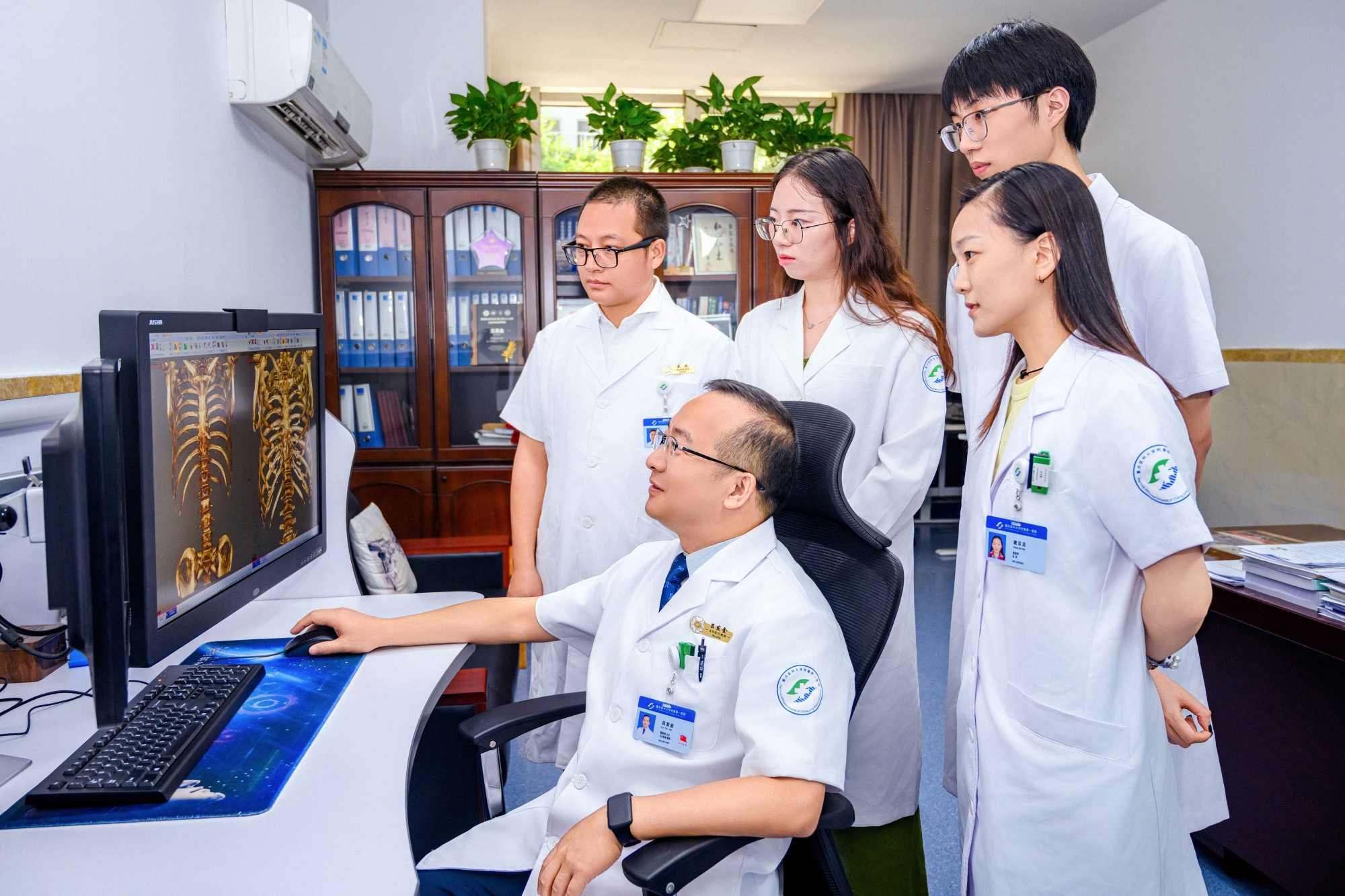
World’s First Research on Bone Calcium CT Imaging Visualization and Quantification Technology
This original, independently patented technology enables precise imaging evaluation of bone quality, early screening for osteoporosis, and three-dimensional visualization and quantitative assessment of pre- and post-treatment changes. In comparison with the current “gold standard” methods for bone density assessment - dual-energy X-ray absorptiometry (DXA) and quantitative CT (QCT) - this technique overcomes limitations related to soft tissue interference, osteophyte formation, and anatomical restrictions, while providing a 3D visual distribution of bone density. Utilizing a single low-dose chest CT scan, it yields four key results: early lung cancer screening, bone density measurement with 3D display, and liver fat content quantification. This method obviates the need for separate equipment and examinations for osteoporosis screening, thereby better fulfilling the demands of public health management and clinical diagnosis and treatment. Initiated in 2022, the technology has already been applied in over 10,000 osteoporosis screenings and more than 1,000 follow-ups after calcium supplementation, resulting in one published SCI paper, one national invention patent, two international conference presentations, two poster presentations, and three additional SCI papers under review.


